| Daylily Spring Sickness |
| Home | Images | Phenology | FAQ | Feedback | Links | What's New |
Diagnostic ImagesClick on any image for a larger picture and use your back button to return here after viewing. 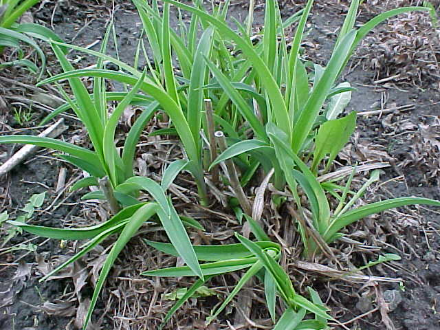
Image 1 shows a common early sign associated with spring sickness, and that is the sideways growth of one or more fans in the clump. You can clearly see the normal growth behind the two affected fans. Not all fans grow sideways to this extent. Many start to curve but their growth then slows down or stops and they remain stunted for varying lengths of time. 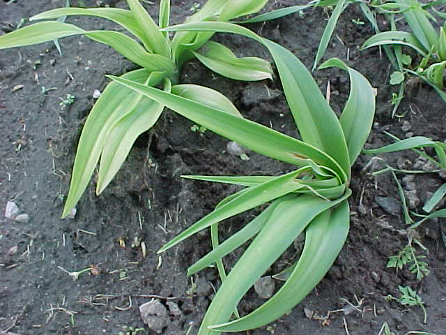
Image 2 shows a fan which has the typical "windswept" look. It has bent sideways but the leaves have not elongated as much as those in the previous image. You can easily see that there are no normal new leaves appearing from the center of the fan. 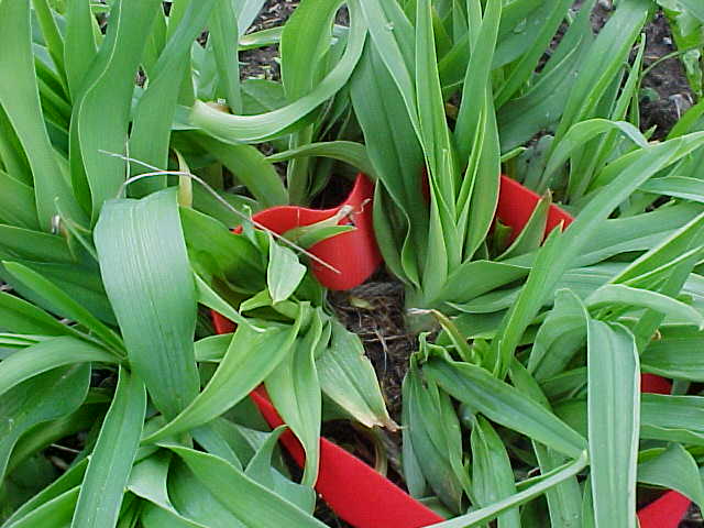
Image 3 shows a clump within which several fans have ceased growing, at least for the time being. These fans are much smaller than the normal ones which surround them. The red ribbon indicates an area of the clump visited the previous fall by a female lesser bulb fly, Eumerus tuberculatus, which was seen to deposit eggs on the old leaves and in soil surface cracks. The fans in the marked area ultimately did develop spring sickness, but we cannot be sure if the bulb fly was involved in the damage or was merely attracted to injuries already in existence. 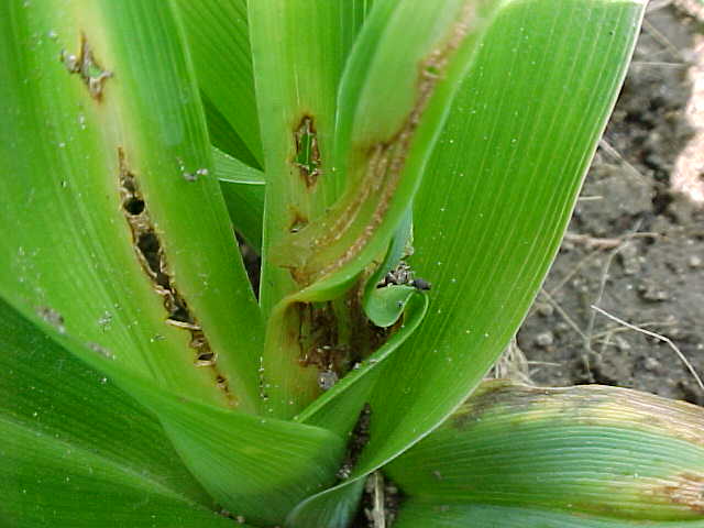
Image 4 shows the typical brown lesions of a daylily fan trying to grow out of spring sickness. This was the only fan in its clump to be affected, the initial sign of trouble being a slight curving of the shoot to one side. Once spring sickness symptoms had become apparent, the other healthy fans in the clump left this one behind in growth. An interesting aspect of this image is the opportunity it gives to compare late stage lesions with the early damage shown in the next picture. 
Image 5 illustrates early stage damage to the innermost baby leaf of a fan (photographed on a one cent coin for size comparison). A fan with spring sickness was examined for clues leaf by leaf until this was the last one left. There is a noticeable similarity between the lesions on the older leaf in the previous image, and those on this younger one. Note the hole with a line of damage extending downwards below it on both specimens. 
Image 6 shows a few struggling fans in front of an otherwise normal clump. The fan on the left has managed more growth but is curving and has some brown lesions. This plant also sustained hail damage so not all of the raggedness in this fan is caused by spring sickness. The fan adjacent to it on the right is bending away from the camera and the leaves are short and rigid compared to normal healthy arching growth. 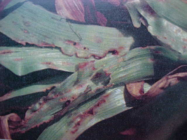
Image 7 is a close up of the brown lesions characteristic of spring sickness. These lesions start off very small and then grow with the plant as illustrated in images 4 and 5. The leaf edges die, break away and crack open giving a sometimes saw-toothed appearance. This is typical of the damage which gives many gardeners the impression that a pest has been chewing on the leaves after spring emergence, but that is not the case. 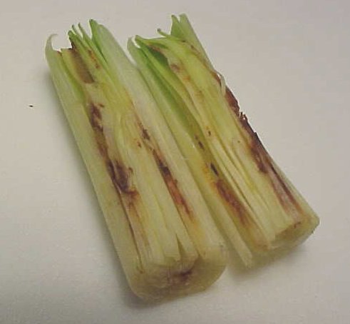
Image 8 - the ragged leaf edges and holes which appear when a fan with spring sickness emerges after growth has commenced in the spring actually start much earlier. This image shows a fan before the damage becomes apparent externally. This fan appeared relatively normal from casual viewing but when cut in half, the extensive internal damage became apparent. 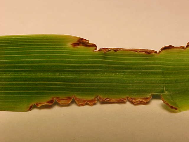
Image 9 The last image in this section shows a close up of the very characteristic "saw-toothed" leaf margins which are so typical of spring sickness. This damage starts much earlier, as shown in the previous image, but as time goes by it dries out and, when the leaf grows, the cracks enlarge and parts of the leaf break off resulting in these ragged edges. |
| View Image Gallery of Spring Sickness and Other Injuries... |
| View Images of Experiments (What's New)...
|
| Home | Images | Phenology | FAQ | Feedback | Links | What's New |
This page last updated April 22nd 2002 ©copyright 2001-2019 Images by Susan Bergeron, except Image 7 by Patricia Henley |